
Fichier:Schistosoma haematobium egg 4843 lores.jpg

Pas de plus haute résolution disponible.
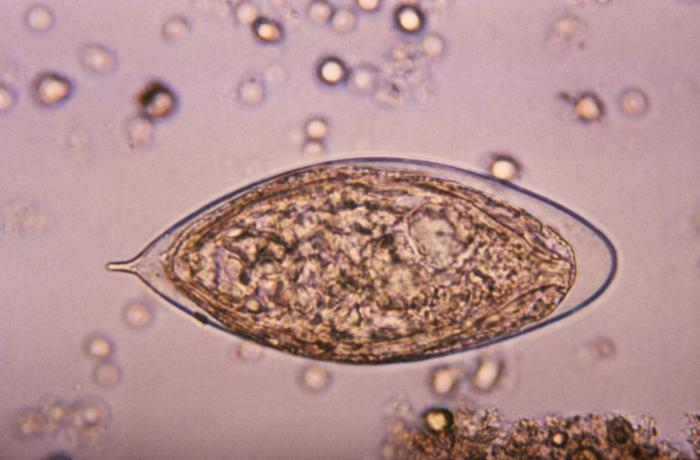
Schistosoma_haematobium_egg_4843_lores.jpg (700 × 460 pixels, taille du fichier : 38 kio, type MIME : image/jpeg)
Ce fichier et sa description proviennent de Wikimedia Commons.
| Description |
ID#:4843 This micrograph depicts an egg from a Schistosoma haematobium trematode parasite; magnified 500x. Note the egg's posteriorly-protruding, terminal spine, unlike the spinal remnant, which protrudes from the lateral wall of the Schistosoma japonicum egg. These eggs are eliminated in an infected human's feces or urine, and under optimal conditions in a watery environment, the eggs hatch and release "miracidia", which then penetrate a specific snail intermediate host. Once inside the host, the S. haematobium parasite passes through two developmental generations of sporocysts, and are released by the snail into its environment as "cercariae". |
|||
| Date | ||||
| Source | http://phil.cdc.gov/PHIL_Images/20031013/b47fc1793d7443d7a5cdbfbc73d95e53/4843_lores.jpg | |||
| Auteur | CDC, Public Health Image Library (PHIL) | |||
| Autorisation (Réutilisation de ce fichier) |
|
Historique du fichier
Cliquer sur une date et heure pour voir le fichier tel qu'il était à ce moment-là.
| Date et heure | Vignette | Dimensions | Utilisateur | Commentaire | |
|---|---|---|---|---|---|
| actuel | 7 mai 2006 à 19:13 | | 700 × 460 (38 kio) | Patho | {{Information| |Description=ID#: 4843 Description: This micrograph depicts an egg from the trematode parasite Schistosoma japonicum with its vestigial spine. The Schistosoma japonicum egg is typically oval or subspherical, has a vestigial spine, and is |
Utilisation du fichier
La page suivante utilise ce fichier :
Usage global du fichier
Les autres wikis suivants utilisent ce fichier :
- Utilisation sur ar.wikipedia.org
- Utilisation sur cs.wikipedia.org
- Utilisation sur de.wikibooks.org
- Utilisation sur en.wikipedia.org
- Utilisation sur gl.wikipedia.org
- Utilisation sur ha.wikipedia.org
- Utilisation sur nl.wikipedia.org
- Utilisation sur sw.wikipedia.org
- Utilisation sur tr.wikipedia.org
- Utilisation sur zh.wikipedia.org