
Fichier:Deep brain stimulation electrode placement reconstruction.png
Taille de cet aperçu : 728 × 600 pixels. Autres résolutions : 291 × 240 pixels | 583 × 480 pixels | 930 × 766 pixels.
Fichier d’origine (930 × 766 pixels, taille du fichier : 904 kio, type MIME : image/png)
Ce fichier et sa description proviennent de Wikimedia Commons.
Description
| Description |
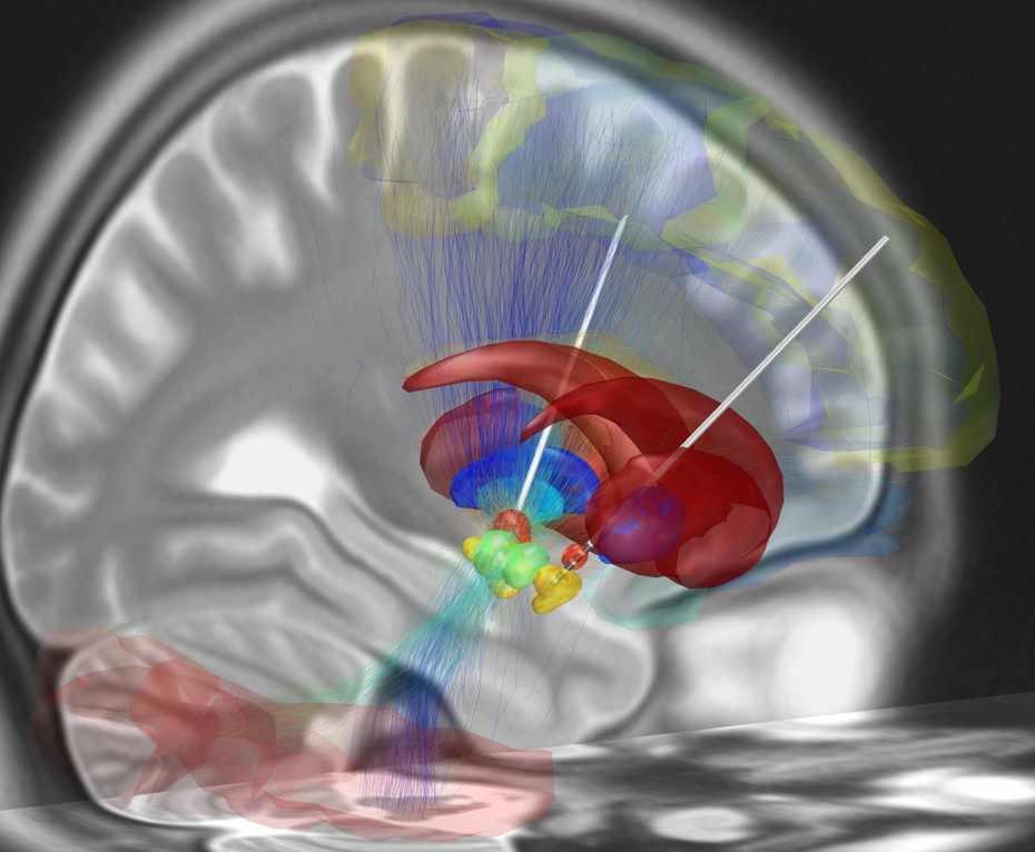
English: Depicted is a reconstruction of bihemispheric DBS electrodes that have been surgically placed into the most common target structure for treatment of Parkinson's Disease, the subthalamic nucleus (orange). Other subcortical structures include the red nucleus (green), the substantia nigra (yellow), the internal (cyan) and external (blue) pallidum and the striatum (red). A stimulation volume is modeled by applying 2V (at 1000Ω impedance) to the second-uppermost contact of the left electrode. Structural fibertracts traversing through this volume are visualized and cortical regions that they connect with the stimulation volume are selected from an automatic anatomical labeling atlas and visualized.
Picture created using LEAD DBS software (www.lead-dbs.org) |
| Date | |
| Source | Travail personnel |
| Auteur | Andreashorn |
Conditions d’utilisation
Moi, en tant que détenteur des droits d’auteur sur cette œuvre, je la publie sous la licence suivante :
Ce fichier est sous la licence Creative Commons Attribution – Partage dans les Mêmes Conditions 4.0 International.
- Vous êtes libre :
- de partager – de copier, distribuer et transmettre cette œuvre
- d’adapter – de modifier cette œuvre
- Sous les conditions suivantes :
- paternité – Vous devez donner les informations appropriées concernant l'auteur, fournir un lien vers la licence et indiquer si des modifications ont été faites. Vous pouvez faire cela par tout moyen raisonnable, mais en aucune façon suggérant que l’auteur vous soutient ou approuve l’utilisation que vous en faites.
- partage à l’identique – Si vous modifiez, transformez, ou vous basez sur cette œuvre, vous devez distribuer votre contribution sous la même licence ou une licence compatible avec celle de l’original.
Historique du fichier
Cliquer sur une date et heure pour voir le fichier tel qu'il était à ce moment-là.
| Date et heure | Vignette | Dimensions | Utilisateur | Commentaire | |
|---|---|---|---|---|---|
| actuel | 16 mai 2015 à 22:06 | | 930 × 766 (904 kio) | Andreashorn | User created page with UploadWizard |
Utilisation du fichier
La page suivante utilise ce fichier :
Usage global du fichier
Les autres wikis suivants utilisent ce fichier :
- Utilisation sur de.wikipedia.org
- Utilisation sur en.wikipedia.org
- Utilisation sur en.wikiversity.org
- Utilisation sur sr.wikipedia.org